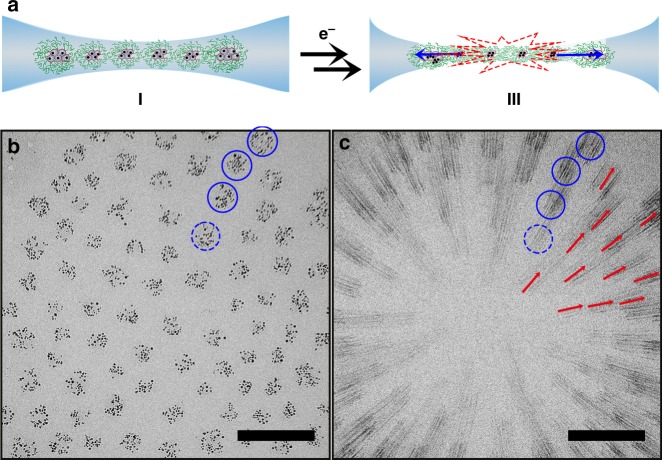
Fig. 2.
Electron beam irradiation-induced rupture of monolayer dendrimicelle superstructures. a Prolonged exposure of G6-Au128-based dendrimicelles results in the rupture of the thin film in which they are embedded. CryoTEM micrographs of the dendrimicelles superstructure before (b) and during the nanoexplosion (c). Because of the relatively long exposure time and the contrast provided by the gold nanoparticles, the rupture process is captured as streaks. The rupture starts from the center of the dendrimicelle superstructures (as indicated with an asterisk) and radially propagates outwards. The blue solid circles are drawn as a guide to the eye around four dendrimicelles before and after rupture. The red arrows are drawn as a guide to the eye to show the directionality of the nanoparticle streaks. Scale bars are 100 nm

