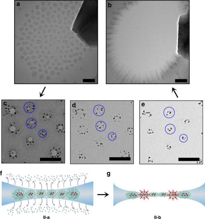
Fig. 4.
Local and global migration in dendrimicelle superstructures under electron beam irradiation. a CryoTEM micrograph of a dendrimicelle superstructure obtained from dendrimicelles encapsulating ninth-generation PAMAM dendrimers in their core, with an Au1024 nanoparticle encapsulated inside every dendrimer. b CryoTEM micrograph after the electron beam-induced nanoexplosion of the dendrimicelle superstructure. c–e Enlarged sections of the superstructure at various time points during the electron beam irradiation, showing that upon prolonged irradiation, the contrast provided by the dendrimicelle core is reduced, and the gold nanoparticles appear closer together. The blue circles in c–e represent the original dendrimicelle core area, and are drawn to visualize the observed dendrimicelle core shrinkage. f, g Schematic illustration of the different stages during the nanoexplosion. CryoTEM sample preparation provides a dendrimicelle superstructure located at the thinnest part of the biconcave water film. f Extensive electron irradiation results in the evaporation of water from the biconcave thin film, leading to the formation of a freestanding polymer film in the center of the grid hole, in which the dendrimicelle cores seems to have shrunk, but more likely the gold particles have been excreted from the dendrimer voids and group together (g). The scale bars are 100 nm

