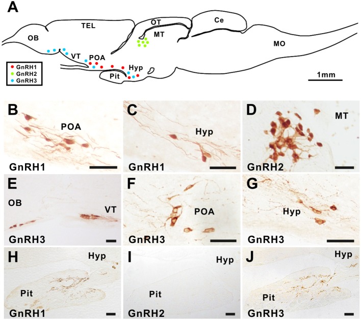
Figure 1.
GnRH1, GnRH2, and GnRH3 immunostaining in the brain and pituitary of ricefield eels. Sagittal sections of the brains together with the pituitary gland of ricefield eels were immunoreacted with primary antibodies, the rabbit polyclonal antibody AS-691 for GnRH1 [1:7,000 dilution; (B,C,H)], the rabbit polyclonal antibody 675 for GnRH2 [1:2,000 dilution; (D,I)], or the mouse monoclonal antibody LRH13 for GnRH3 [1:2,000 dilution; (E–G,J)]. After incubation with primary antibodies for 40 h at 4°C, sections were then exposed to the secondary antibody, HRP-conjugated goat anti-rabbit or anti-mouse IgG (H+L) (1:1,000 dilution; Beyotime, Shanghai, China), and finally visualized with 3,3'-diaminobenzidine (DAB) solution, mounted, and digitally photographed with a Nikon Eclipse Ni-U microscope (Japan). (A), the schematic diagram of the ricefield eel brain and pituitary. Red, green, and blue dots in (A) represent GnRH1, GnRH2, and GnRH3 immunoreactive neurons, respectively. OB, olfactory bulbs; TEL, telencephalon; VT, ventral telencephalon; POA, preoptic area; OT, optic tectum-thalamus; MT, midbrain tegmentum; Hyp, hypothalamus; Pit, pituitary; Ce, cerebellum; MO, medulla oblongata. Scale bar = 50 μm except where it is specifically designated otherwise.

