Figure 3.
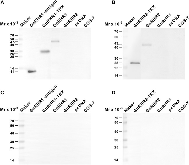
Specificities of anti-GnRHR1 and anti-GnRHR2 antisera against recombinant proteins as determined by Western blot analysis. The recombinant proteins (50 ng) or COS-7 cell extracts (100 μg) were separated on 12% SDS-PAGE gels, processed routinely, and immnoreacted with the rabbit anti-GnRHR1 antiserum [1:2,000 dilution; (A)], the mouse anti-GnRHR2 antiserum [1:1,000 dilution; (B)], the anti-GnRHR1 antiserum pre-adsorbed with 10 ug/mL of recombinant GnRHR1 expressed in transfected COS-7 cells (C), or the anti-GnRHR2 antiserum pre-adsorbed with 10 ug/mL of recombinant GnRHR2 expressed in transfected COS-7 cells (D). The secondary antibody was 1:1,000 diluted horseradish peroxidase (HRP)-conjugated goat anti-rabbit or anti-mouse IgG (H+L) (Beyotime, Shanghai, China), and the blots were visualized using BeyoECL Plus kit (Beyotime). GnRHR1-antigen, the recombinant GnRHR1 polypeptide used to immunize rabbit; GnRHR1-TRX, the region corresponding to GnRHR1 antigen expressed as thioredoxin (TRX) fusion protein; GnRHR2-TRX, GnRHR2 polypeptide region encompassing the synthetic antigen peptide expressed as TRX fusion protein. GnRHR1 and GnRHR2, the recombinant full-length ricefield eel GnRHR1 and GnRHR2 proteins expressed in transiently transfected COS-7 cells. pcDNA, extracts of COS-7 cells transfected with the empty vector pcDNA3.0. COS-7: extracts of COS-7 cells.

