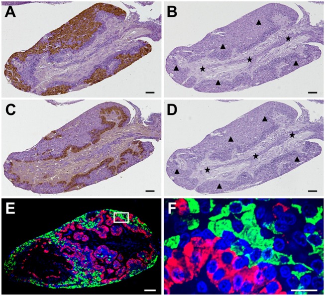
Figure 4.

Specificities of GnRHR1 and GnRHR2 immunoreactivities in the pituitary of male ricefield eels as determined by immunohistochemical analysis. Sagittal sections of the pituitary gland were immunoreacted with the rabbit anti-GnRHR1 antiserum [1:500 dilution; (A)], the pre-adsorbed anti-GnRHR1 antiserum by 10 ug/mL of recombinant GnRHR1 expressed in transfected COS-7 cells (B), the mouse anti-GnRHR2 antiserum [1:500 dilution; (C)], the pre-adsorbed anti-GnRHR2 antiserum by 10 ug/mL of recombinant GnRHR2 expressed in transfected COS-7 cells (D), or the mixture of the rabbit anti-GnRHR1 (1:500 dilution) with the mouse anti-GnRHR2 (1:500 dilution) antisera (E,F). The secondary antibody was 1:1,000 diluted horseradish peroxidase (HRP)-conjugated goat anti-rabbit or anti-mouse IgG (H+L) (Beyotime, Shanghai, China) for (A–D), and the mixture of Alexa Flour 488-labeled goat anti-rabbit IgG (H+L) (1:500 dilution) and Cy3-labeled goat anti-mouse IgG (H+L) (1:500 dilution) for (E). DAPI was used to stain the nuclei blue. Immunostaining (brown) in (A–D) was visualized by the DAB chromogen and counterstained with hematoxylin. The black triangles and stars indicate the adenohypophysis and neurohypophysis tissues, respectively. The image of (E) was captured and analyzed with a Nikon i-E confocal microscope equipped with a CSU-W1 spinning-disk head (Yokogawa, Tokyo, Japan) for immunofluorescent staining of GnRHR1 (green) and GnRHR2 (red). (F) is the higher magnification of the boxed area in (E). Sagittal sections of ricefield eel pituitary glands were shown here with the rostral (anterior) to the left. Scale bar = 50 μm except the image of (F) was 10 μm.
