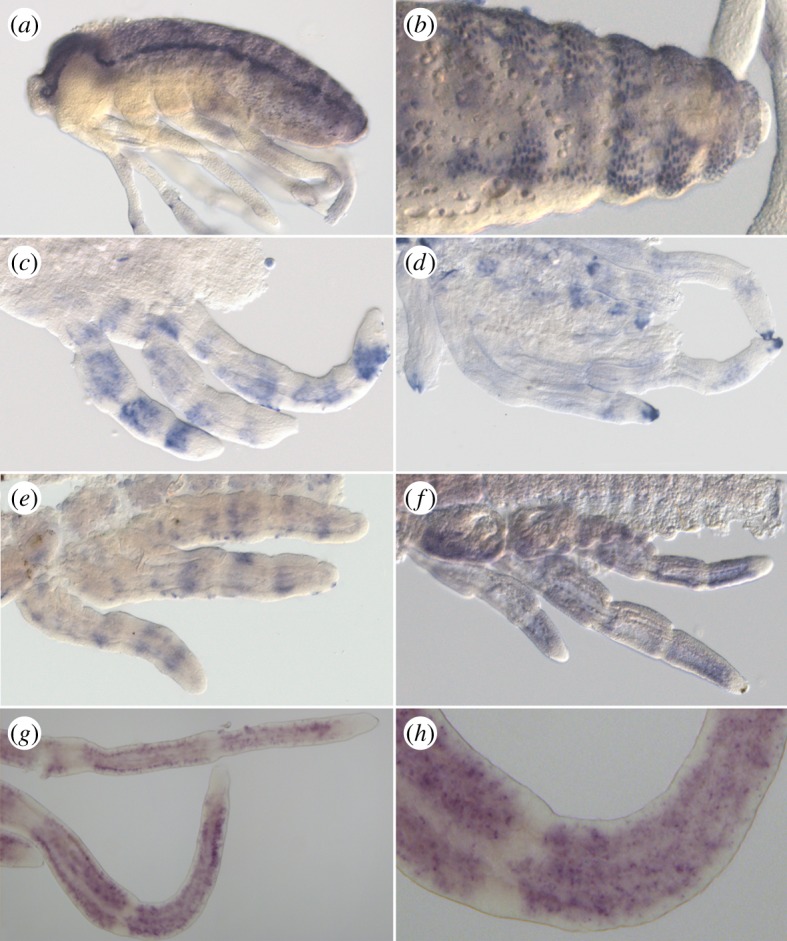
Figure 5.

In situ hybridization showing ASH expression pattern. (a,b) Staining of ASH in Mesovelia mulsanti. ASH is expressed during central nervous system development in ectodermal cell clusters (a) that are progressively restricted to neural precursors (b). (c,d) Staining of ASH in Microvelia americana. In post-katatrepsis embryos, ASH is expressed in discrete large transverse bands along the leg (c). This staining is restricted to the distal tip of the tarsus in later stages (d). Staining of ASH in Gerris buenoi (e–h). Similarly to Mi. americana embryos, ASH expression pattern is dynamic in legs in sharp transverse bands (e), in larger bands (f), then in spotty domains (g,h).
