FIGURE 1.

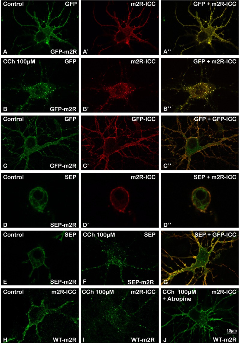
Validation of GFP-m2R, SEP-m2R, and WT-m2R expressing vectors and localization of transfected m2R in hippocampal neurons. Hippocampal neurons were transfected with a plasmid encoding the GFP-tagged receptor (GFP-m2R) (A–C”), SEP-tagged receptor (SEP-m2R) (D–G) or wild-type receptor (WT-m2R: H–J), fixed, and processed for visualization of the receptor by confocal microscopy. Equatorial images of neurons (0.5 μM in depth) were selected and illustrated on this panel. The m2R localization was identified by GFP (A,A”,B,B”,C,C”) or SEP (D,D”E,F,G) native fluorescence or by fluorescent ICC using an anti-m2R (m2R-ICC : A’,B’,D’,H–J) or anti-GFP antibody (GFP-ICC : C’,G). Whatever the construction, the fluorescent signal is localized at the membrane of the soma and the dendrites. A faint signal is detected in the cytoplasm. (B,F,I) The stimulation with a muscarinic receptor agonist [Carbachol (CCh), 100 μM] induces a huge decrease of the signal at the somatic and dendritic membranes and a the appearance of a punctiform labeling in the cytoplasm when using a GFP (B) or SEP-tagged (F) or WT (I) construction. (J) A neuron, that has been pre-incubated with Atropine (10 nM), a muscarinic receptor antagonist, display a membrane labeling at the soma and dendrites similar to a control staining. (A–B”,D–D”) Fluorescent signals detected by direct visualization of GFP (A,B) or SEP (D), and by m2R ICC (A’,B’,D’) in a same neuron perfectly colocalize (A”,B”,D”). (C–C”) GFP or SEP detection by ICC (C’,G) display a membrane labeling in a non-permeabilized neuron that colocalizes with the direct GFP or SEP fluorescence (C,C”,G).
