
FIGURE 1.
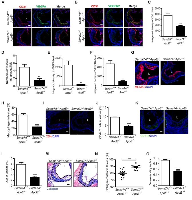
Sema7A deletion reduces intraplaque neovascularization via VEGFA/VEGFR2 downregulation and impairing inflammatory cell infiltration and remarkably enhances plaque stability in ApoE-/- mice. (A,B) Aortic root sections from Sema7A+/+ApoE-/- and Sema7A-/-ApoE-/- mice on HFD for 12 weeks. Endothelial cells were stained with CD31 and VEGFA/VEGFR2 (CD31, red; VEGFA/VEGFR2, green; nuclei, blue). Data are shown as mean ± SEM (n ≥ 10 mice per group). Bar = 100 μm. (C,E,F) Integrated density of CD31/VEGFA/VEGFR2 was qualified from (A,B). Data are shown as mean ± SEM (n ≥ 10 mice per group). Bar = 100 μm, ∗∗P < 0.01; ∗∗∗P < 0.001. (D) Quantified data for numbers of vessels intraplaque/filed. Data are shown as mean ± SEM (n ≥ 10 mice per group). Bar = 100 μm, ∗∗P < 0.01. (G–L) Aortic root sections were also stained for macrophages (G, MOMA-2, red; nuclei, blue), T cells (I, CD4, red; nuclei, blue) and DCs (K, CD11c, green; nuclei, blue), Data are shown as mean ± SEM (n ≥ 10 mice per group). Bar = 100 μm. ∗∗∗∗P < 0.0001. (M) Collagen content (blue) in aortic root sections was stained with Masson’s Trichrome and nucleus was counterstained with hematoxylin. L, aortic lumen. (N) The result was shown as percent of the positive area in plaque area. Data are shown as mean ± SEM. n ≥ 10 mice per group. Bar = 200 μm. ∗∗∗P < 0.001. (O) The vulnerability index, an indicator of plaque stability, in Seme7A+/+ApoE-/- and Seme7A+/+ApoE-/- was calculated. Vulnerability index = (macrophage content % + lipid content %)/(collagen content % + VSMC content %) Data are shown as mean ± SEM, n ≥ 10, ∗∗∗∗P < 0.0001.