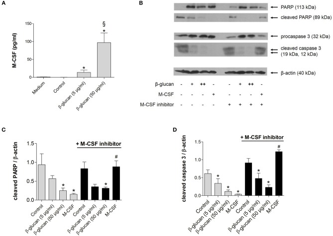
Figure 2.
The anti-apoptotic effect of β-glucan is independent of M-CSF. (A) Human monocytes were left untreated (control) or stimulated for 24 h with β-glucan (5 μg/ml or 50 μg/ml). The release of M-CSF was determined by cytometric bead array in cell supernatants or in medium plus 10% serum only (medium). (B–D) Human monocytes were preincubated with the M-CSF inhibitor GW2580 or vehicle for 60 min and then incubated for 48 h with either M-CSF (50 ng/ml) or β-glucan (5 μg/ml or 50 μg/ml). Controls were left without stimulation. Protein lysates were analyzed in immunoblots for cleavage of PARP and caspase 3. (B) One representative blot is shown. (C,D) Densitometry analyses of protein bands normalized to β-actin of three independent experiments. Values are means ± SEM, *p < 0.05 compared to the respective untreated control, §p < 0.05 compared to 5 μg/ml β-glucan, #p < 0.05 compared to M-CSF alone.

