Figure 1.
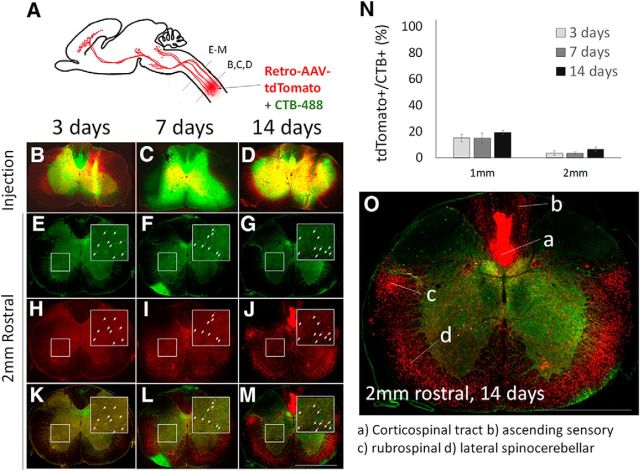
AAV2-Retro shows limited retrograde transduction of cervical propriospinal neurons. A, Mixed AAV2-Retro-tdTomato and CTB-488 were injected bilaterally to C4/5 gray matter, and transverse spinal sections at the injury site and 1 or 2 mm rostral were examined 3, 7, or 14 d later. B–D, Transverse sections at the level of spinal injection show readily detectable CTB-488 and tdTomato. E–G, In cervical spinal cord 2 mm rostral to the injection, CTB-488 retrogradely labels cell bodies that project axons to the injection site (arrows). H–M, tdTomato signal is rarely detectable in CTB-488+ cell bodies, indicating low transduction. N, Quantification of tdTomato detection in CTB-488+ cell bodies showed that <20% of propriospinal neurons projecting to C4/5 were transduced 1 mm rostral to the injury, and <10% at 2 mm. O, By 14 d postinjection, tdTomato signal is apparent in locations corresponding to ascending and descending axon tracts. N > 100 individual cells from each of at least four animals at each time point. Error bars show SEM. Scale bars, 1 mm. Statistical comparisons of transduction efficiencies between supraspinal populations are provided in Figure 1-1.

