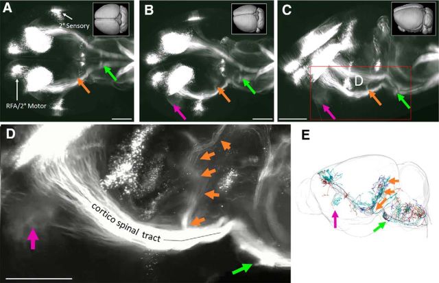
Figure 6.
AAV2-Retro and 3D imaging reveal CST cell body distribution, axon trajectories, and collateralization. A–D, Adult mice received bilateral cervical injection of AAV2-Retro-tdTomato. Two weeks later brains were optically cleared by 3DISCO and imaged with light-sheet microscopy. Images showed rotated views of whole brain, with rostral to the left and initially viewed from the dorsal surface in A. Three distinct groups of CST cell bodies are apparent, and to clear points of collateral branching from the main CST are visible (arrows). E, Individual tracings of CST neurons from the Mouselight Neuron Browser. Consistent with the 3D reconstruction, collateral branches to tectal areas (orange arrow) and to Basilar Pontine Nuclei (green arrow) are visible. Videos of cleared brains are available in Movies 1, 2, 3, and 4. Scale bars, 1 mm.

