
Fig. 1.
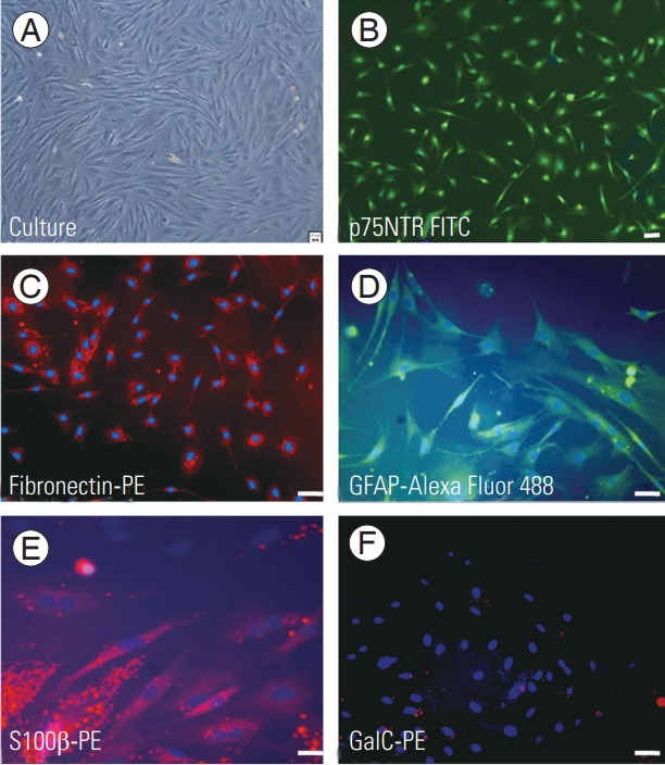
Immunohistochemical characterization of OECs and ONFs. Image showing culture in phase contrast (A), OECs stained with P75NTR-fluorescein isothiocyanate (B), and ONF positively stained with fibronectin-PE (C). OEC/ONF stained with GFAP-Alexa Fluor 488 (D) and S100β-PE (E) showing positivity for astrocyte and Schwann cell markers. (F) OEC/ONF stained with GalC-PE showing negativity for the marker. Scale bar=20 μm. OEC, olfactory ensheathing cell; ONF, olfactory nerve fibroblast; PE, phycoerythrin.