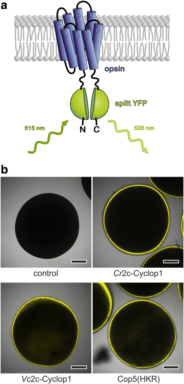
Fig. 2.

2c-Cyclops possess cytosolic N-termini and a likely 8 transmembrane helices topology. a Schematic model of BiFC (bimolecular fluorescence complementation) experiments. The opsin domain was N- and C-terminally fused to the two parts of split YFP (YFPC = aa155–238 of YFP, YFPN = aa1–154 of YFP). b Fluorescence pictures show the following: control oocyte (control), oocytes expressing YFPC::Cr2c-Cyclop1/opsin::YFPN (Cr2c-Cyclop1), YFPC::Vc2c-Cyclop1/opsin::YFPN (Vc2c-Cyclop1), and YFPC::Cop5/opsin::YFPN (Cop5(HKR)) constructs. The fusion sequence of YFPC and YFPN was designed according to the YFP structure to facilitate the fluorescence complementation. The fluorescence images were taken by a confocal microscope 3 dpi (days post injection) with 30 ng cRNA injection into Xenopus oocytes. Scale bars 250 μm
