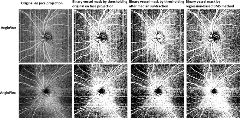
Figure 4.
Comparison of vascular maps of the nerve fiber layer plexus en face projection of a representative scan of a healthy subject generated by thresholding the original scan without BMS, median subtraction, and the regression-based BMS algorithm. Scans acquired by both commercial systems used in this study are represented. Segmentation of the image corresponding to Optovue's BMS method is different because it was done on a different file, generated by RTVue software.

