
Figure 6.
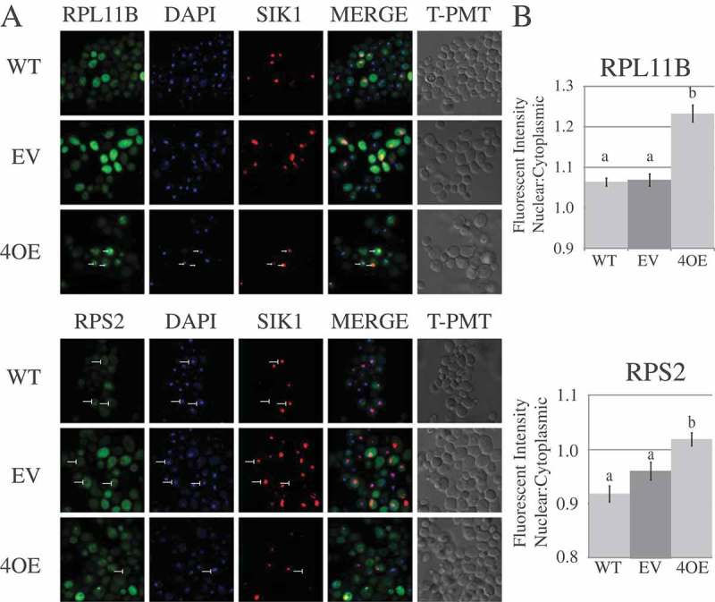
Puf4p overexpression results in nuclear accumulation of ribosomal subunits. (a) Representative panels are shown from confocal microscopy of cells from the WT strain, or the WT strain containing an empty vector (EV) or a vector overexpressing Puf4p (4OE). Labels above the panels indicate the stain/protein being observed. Arrows on the upper set of panels indicate the position of nuclear foci as detected by RPL11B-eGFP. Arrows on the lower set panels indicate the position of nuclei in the cells as detected by a decreased signal of RPS2-eGFP. (b) Graphical representation of the nuclear to cytoplasmic ratio of GFP fluorescence intensity in WT cells ± empty vector or vector overexpressing Puf4p. Letters a and b indicate significant differences using one-way ANOVA (p < 0.005) and Tukey’s post-hoc test. Error bars represent SEM and are representative of ≥ 250 cells spread across 3 individual growth trials.