
Figure 9.
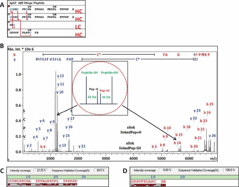
MS/MS spectrum identifying the IgG2 A/B specific hinge peptide (m/z 6886.253) with fragment ions assigned (B). The structure of the asymmetric peptide with 5 disulfide bonds is shown in (A). Bottom: the fragment ion coverage of the hinge peptide (C) and the linked HC 12mer peptide (D) are shown. The peak quartet at m/z 1230.630 corresponds to the 12mer HC peptide and the quartet at m/z 5658.65 to hinge peptide with loss of the LC tripeptide (peak quartets are indicated by red arrows).