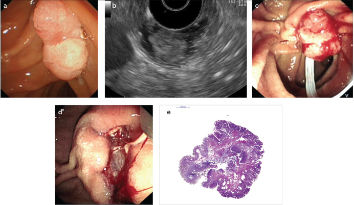
Figure 1. a–e.
Endoscopic view of an ampullary lesion without worrisome features for malignancy (a); endosonographically, the hypoechoic mucosal lesion is not invading into the muscularis mucosa (b); ampullary lesion is grasped with a polypectomy snare for resection (c); post-papillectomy view with a guide-wire advanced into the pancreatic duct prior to pancreatic duct stent placement (d); histopathologic evaluation (H&E stain) revealed a tubulovillous adenoma with high-grade dysplasia foci and negative margins (e)

