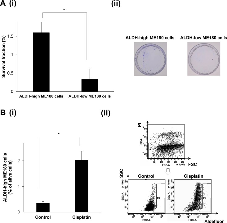
Figure 2. The radio- or chemoresistant nature of the ALDH-high ME180 cells.
(A) Radioresistant nature of the ALDH-high ME180 cells assessed by clonogenic survival assay. ALDH-high and ALDH-low ME180 cells (1 × 10³) were severally plated in 60 mm dishes and treated with 4 Gy of radiotherapy in the presence of 10% of FBS. As a control group, 100 cells of ALDH-high and ALDH-low ME180 cells were plated. After they had been cultured for 3 weeks, the colonies were stained with 0.5% crystal violet and the numbers of colonies were counted. The survival fractions (SF) were calculated by following formulas. SF = 100 × plating efficacy (PE) of treated sample/PE of control. PE = number of colonies/number of cells plated. (i) The survival fractions of ALDH-high and ALDH-low ME180 cells (Bars SD. n = 3, p < 0.01, two-sided Student’s t test). (ii) Representative photos of the colonies formed by the ALDH-high and ALDH-low cells treated with 4 Gy of radiotherapy. (B) Chemoresistant nature of the ALDH-high ME180 cells. ME180 cells (3 × 10⁶) were inoculated with 1 mM of cisplatin or PBS (control) in the presence of 10% of FBS for 3 days in 100 mm dishes. Among the surviving ME180 cells (propidium iodide (PI)-negative cells), the percentages of ALDH-high cells were assessed using the Aldefluor assay. (i) The percentage of ALDH-high ME180 cells (Bars SD. n = 4, p < 0.05, two-sided Student’s t test). (ii) Representative dot plots are shown.

