Figure 3.
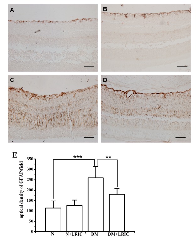
LRIC treatment ameliorated retinal Müller cell activation in diabetic rats. There was a significant increase in the level of GFAP expression in the diabetic retina (C) compared with the control retina (A). After 12 weeks of LRIC treatment, GFAP immunostaining decreased significantly in the diabetic retina (D). However, GFAP expression in the control retina was not affected by LRIC (B). Scale bar=50 μm. (E) Bar graphs depicting the the density of GFAP in each group. Data are expressed as mean±SD, ** P<0.01, *** P<0.001. N=5 each group.

