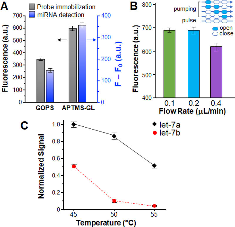
Figure 3. Optimization of the MERCA assay with let-7a miRNA.
(A) Effects of conjugation chemistries on surface immobilization of capture probes (grey) and miRNA detection (blue). To assess probe immobilization, FAM labeled capture probe (50 μM) is covalently immobilized on glass slides by the APTMS-GL and GOPS methods, respectively. For RCA detection, let7a (5.0 × 10⁻¹⁰ M) was measured as a model sample using chip surfaces modified by the two methods. F: fluorescence intensity for miRNA detection; F0: background signal obtained with buffer. (B) Comparison of the detection of let-7a (5.0×10⁻¹⁰ M) using different averaged flow rates for solid-phase miRNA capture. A five-step pumping sequence was used with the actuation time for each step varied from 200 to 500 ms and the pulse time fixed at 2 s (inset). (C) Effects of capture temperature on the specificity of miRNA detection. The concentrations of let-7a and let-7b are 5.0×10⁻¹⁰ M, SYBR Green as fluorescence reagent, respectively. Error bars indicate one standard deviation of three replicates.

