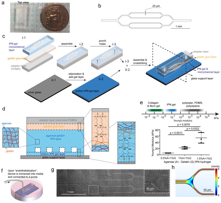
Figure 1. Engineering an interpenetrating network (IPN) hydrogel-based microvasculature-on-a-chip for investigating endothelial barrier function and cellular interactions in hematologic diseases.
a) Macroscopic top view of the microdevice. The dashed square indicates the transparent bottom layer. b) CAD design for the photolithography mask that defines the microfluidic channel geometry. c) Schematic of microdevice fabrication. d) Schematic of agarose-gelatin IPN layer and the bonding of each layers via gelatin using carbodiimide crosslinker chemistry. e) The IPN hydrogel can be tuned to mimic the physiological stiffness of the blood vessel intima, while synthetic polymeric materials are much stiffer. Data were plotted as the mean ± s.d. with n=3 independent experiments. P-values were calculated using unpaired, two-sided Student’s t-test. (* P<0.05, ** P<0.01). f) The microdevice seeded with endothelial cells was immersed into culture media, and maintained under constant physiologic laminar flow for up to 4 weeks. g) A representative stitched composite of brightfield microscopy images of a microvasculature-on-a-chip after 14 days of culture. Insert: higher magnification image of the dashed box. h) Computational fluid dynamics modeling of the microchannels confirming physiologic laminar flow conditions.

