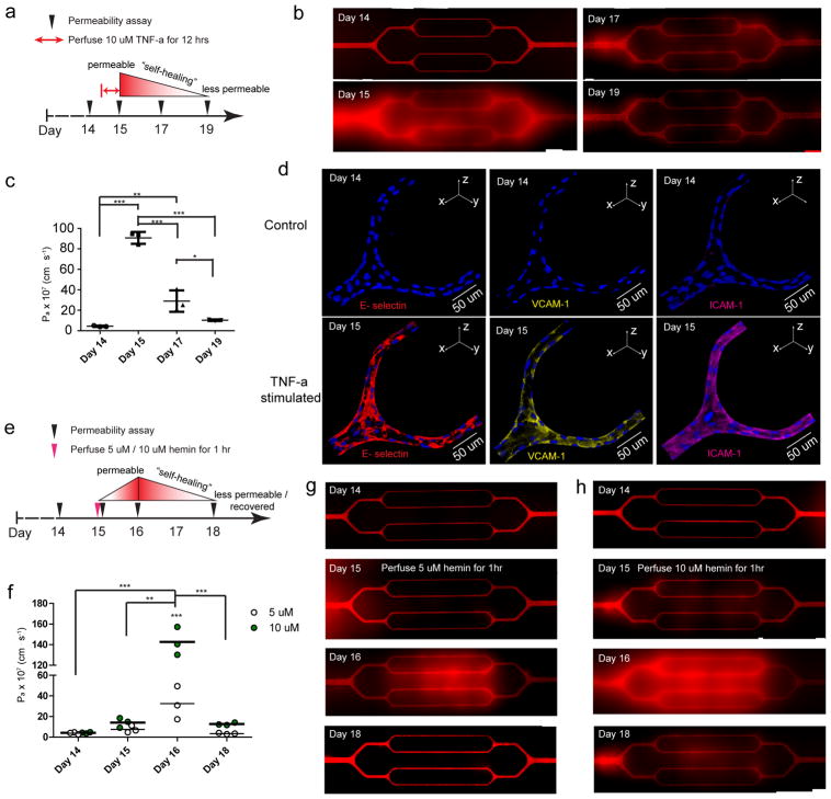
Figure 3. The spatiotemporal dynamics of endothelial barrier dysfunction in response to perfusion of inflammatory cytokines and hemolytic byproducts and the “self-healing” of engineered endothelial barrier integrity upon removal of those agents can be visualized and tracked.
a) The experimental timeline and design with TNF-α. b) Stitched composite of epifluorescence images indicate that exposure to 10 ng/ml TNF-α for 12 hours significantly increased permeability in the engineered microvasculature, but over 4 days, the TNF-α-stimulated endothelium “self-healed” and gradually became less permeable, recovering its barrier function. c) Quantitative apparent permeability measurements of the BSA tracer indicates that perfusion and stimulation with 10 ng/ml TNF-α for 12 hours appropriately and expectedly increased permeability of the engineered microvasculature by 20-fold. Post-TNF-α stimulation, the endothelial barrier function of engineered microvasculature “self-healed” and recovered within 4 days. Data was plotted as the mean ± s.d. with n=3 independent biological replicates. P-values were calculated using one-way ANOVA with Bonferroni’s post hoc test (* P<0.05, ** P<0.01; *** P<0.001). d) Immunostaining indicates that TNF-α stimulation appropriately upregulated the expression of adhesion molecules in the engineered microvasculature. e) The experimental timeline and design with free hemin. f) Quantitative apparent permeability measurements of the BSA tracer at different time points of both pre- and post- hemin exposure indicates that the magnitude of barrier function loss and the system’s “self-healing” recovery thereof were hemin dose-dependent. Data was plotted as the mean ± s.d. with n=3 independent biological replicates. P-values were calculated using two-way ANOVA with Bonferroni’s post hoc test (** P<0.01; *** P<0.001). g–h) Stitched composite of epifluorescence images from permeability measurements at different time points of both pre- and post- exposure to 5 μM and 10 μM hemin.

