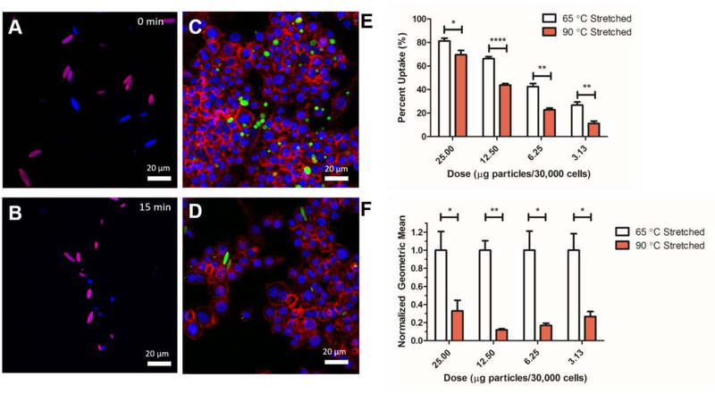
Figure 4.
Phagocytic cells demonstrate different responses to differentially stretched particles that are triggered by the laser. Confocal images of a mixed population of bulk microparticles heated at 45 °C for (A) 0 min and (B) 15 min. For full time course, see Figure S8. 65 °C stretched particles (blue) demonstrate full reversion over the heating whereas 90 °C stretched particles (magenta) demonstrate no reversion to their spherical form. (C) 65 °C stretched and (D) 90 °C stretched particles were cultured with macrophages first heated at 42 °C for 15 min to trigger SME and then incubated at 37 °C for 4 hours. Confocal imaging demonstrates that there is a preference of macrophages to phagocytically take up spherical particles in high quantities compared to non-spherical particles. Blue = DAPI, Red = Actin, Green = Particles. (E) Percent positive uptake and (F) particle fluorescence geometric mean as analyzed by flow cytometry demonstrates that the 65 °C stretched laser triggered shape memory particles were taken up at a higher percentage of the course of 4 hours. Error bars are standard error of n = 4 replicates.

