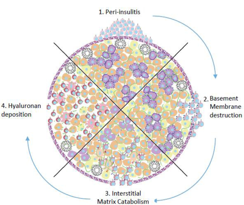
Figure 3: A schematic of the sequential loss of ECM barriers against in autoimmune insulitis.

In healthy islets, the ECM provides physical and immunoregulatory barriers against immune infiltration. However, in autoimmune diabetes, effector T cells evade central and peripheral tolerance and accumulate around islets (top). These auto-reactive cells require a break in the laminin rich basement membrane for entry (right). Degradation of islet HS leads to beta cell death (bottom). Deposition of HA allows for further infiltration, effector cell activation, and inhibition of Treg expansion (left). Along with effects on infiltrating leukocytes, this remodeling of the islet ECM also adversely impacts β-cell health.
