Fig. 3.
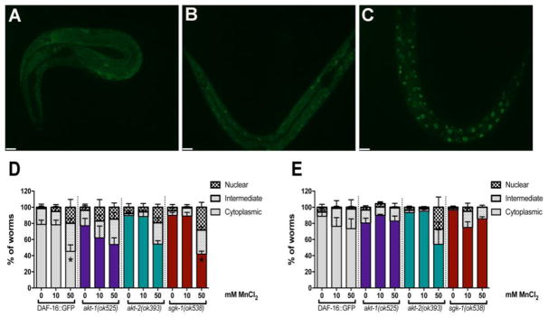
Mn induces DAF-16 nuclear translocation. Akt-1(ok525), akt-2(ok593), sgk-1(ok538) were crossed with Pdaf-16A::GFP. Worms were exposed to 10 or 50 mM MnCl2 for 1 h. Controls were incubated with 85 mM NaCl. Worms were scored as having cytoplasmic (A), intermediate (B) or nuclear (C) DAF-16 distribution. DAF-16 was visualized by fluorescent microscopy within 10 min of Mn exposure (D), or at the end of 1 h exposure (E). Results are expressed as percent of worms with each condition. Data are expressed as mean + S.E.M. from five independent experiments. Statistical analysis by two-way ANOVA followed by post hoc Tukey’s test. Representative images were obtained with a PerkinElmer spinning disk confocal, 40X objective. Scale bar represents 10μm.

