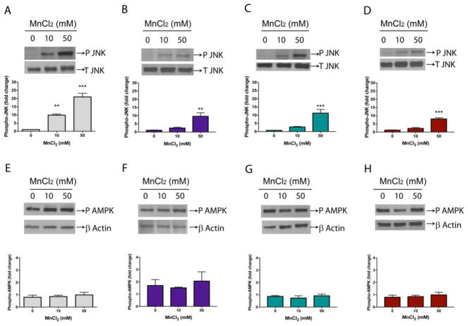
Fig. 7.
Phosphorylated proteins were evaluated by western blotting. Worms were exposed to Mn for 1 h (10 or 50 mM). Samples for SDS-PAGE were prepared immediately after treatment. Blots were developed by chemiluminescence. Bands were quantified by densitometry with imageJ software. (A) Phosphorylation of JNK in wild-type N2 or null mutants (B) akt-1(ok525), (C) akt-2(ok593), (D) sgk-1(ok538). Phosphorylated (P) JNK was normalized to total (T) JNK. (E) Phosphorylation of AAK/AMPK in wild-type N2 or null mutants (F) akt-1(ok525), (G) akt-2(ok593), (H) sgk-1(ok538). P AMPK was normalized to β-actin. Data represent fold change compared to controls and express the mean ± S.E.M. of 4 experiments. ** p < 0.01, *** p < 0.001 compared to controls (one-way ANOVA followed by post hoc Tukey’s test).

