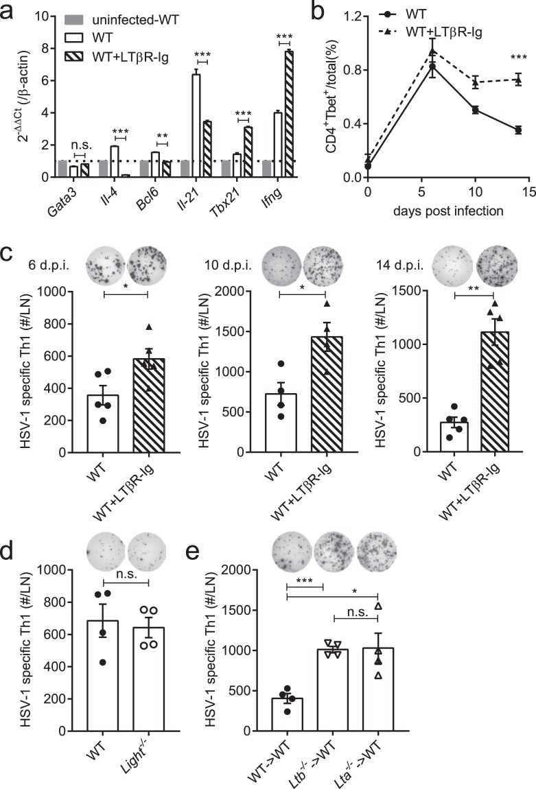
Figure 1.
LT-LTβR signaling deficiency enhances the anti-HSV-1 Th1 response. (a) Expression of the Th1/Th2/Tfh-related transcriptional factors and cytokines in purified CD4+ T cells on day 4 p.i. in the popliteal LNs tested by real-time qPCR (n = 3/group). (b) Percentages of CD4+Tbet+ cells in total cells in the draining LNs (popliteal LN and inguinal LN) from WT mice (solid line) and LTβR-Ig-treated mice (dotted line) (n = 5/group). (c) The Th1 response of LTβR-Ig-treated mice on 6 d.p.i. (n = 5/group), 10 d.p.i. (n = 4/group) and 14 d.p.i. (n = 5/group), including immunospots and absolute numbers of IFNγ-secreting CD4+ cells per LN. (d) The Th1 response of Light−/− mice on day 14 p.i. (n = 4/group), including immunospots and absolute numbers of IFNγ-secreting CD4+ cells per LN. (e) The Th1 response of Lta−/− → WT and Ltb−/− → WT bone marrow chimeric mice on day 14 p.i. (n = 4/group), including immunospots and absolute numbers of IFNγ-secreting CD4+ cells per LN. Data are representative of three independent experiments, shown as mean ± SEM. n.s., not significant; *P < 0.05; **P < 0.01; ***P < 0.001. (Two-way ANOVA multiple comparisons for b).

