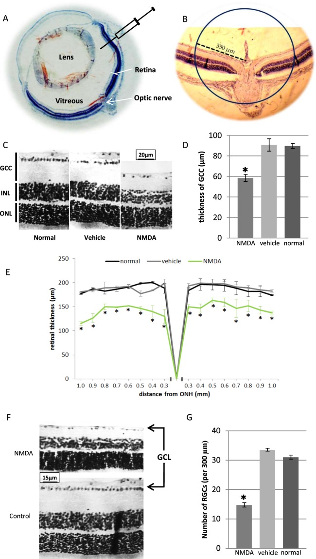
Figure 1.
Generation of mouse experimental model by NMDA excitotoxic amino acid. (A) Schematic illustration of intravitreal injection of NMDA; All injections were performed under ora serrata by glass needle. (B) For evaluation of mice model, analyses were performed on a distinct distance of optic nerve. (C,D) Histological analyses of GCC thickness 7 days post injection. Data significantly decreased in NMDA samples versus vehicle treated and normal mice. (E) Quantitative spider plot analysis depicting significant decrease in retinal thickness in NMDA samples. (F,G) Histological analysis of number of ganglion cells 7 days post injection. Data significantly decreased in NMDA samples versus vehicle treated and normal mice. GCC: ganglion cell complex; ONL: outer nuclear layer; INL: inner nuclear layer; GCL: ganglion cell layer; ONH: optic nerve head. (*P < 0.05 versus other groups, error bar: means ± SD).

