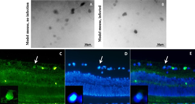
Figure 5.
Analysis of Ki67 expression following Pax6 overexpression that was mediated by AAV2 viruses in model mouse retina. (A) Immunofluorescence of Ki67 in control flatmounts (only NMDA injection, no viruses), 30 days post infection; Ki67 was expressed obviously after NMDA injection. (B) Immunofluorescence of Ki67 in flatmounts, 30 days post NMDA and viruses injection; Ki67 was expressed as the same as the controls. (C–E) Immunostaining of Ki67 on 5 µm section following injections. Ki67 was expressed in GCL and some cells in INL.

