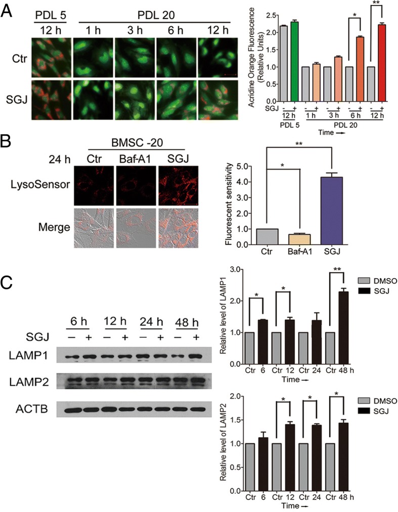
Fig. 2.

SGJ increased the concentration of H+ in lysosomes, and up-regulated LAMP1 and LAMP2 protein level. a Acridine orange staining for young (PDL 5) and senescent (PDL 20) BMSCs. Acidic vacuoles declined with age as shown in the results. Twenty-micromolar SGJ treatments for 1, 3, 6, and 12 h significantly restored the amount of acidic vacuoles (magnification × 200). b SGJ promoted lysosomal acidification. Lysosensor™ Green DND-189 was used to sense the changes of the concentration of H+ in lysosomes, and quantification. BMSCs were treated with 20 nM Baf-A1 or 20 μM SGJ for 24 h. The changes of the red fluorescence reflect changes in lysosomal pH. c Western blot analysis of LAMP1 and LAMP2 protein levels with β-actin as a loading control, and quantification. BMSCs were treated with 20 μM SGJ for 6, 12, 24 and 48 h. (*, p < 0.05; **, p < 0.01, results were expressed as means ± SEM, n = 3)
