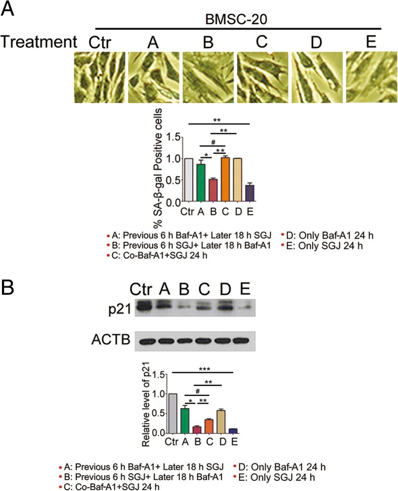
Fig. 5.

Changes in SA-β-gal positive cells and p21 protein level after treatment with SGJ and/or Baf-A1. a BMSCs were treated as the above condition. Senescent cells were stained blue under a phase-contrast microscopy. The percentage of positively stained cells was estimated by counting at least 1500 cells for each sample. b Western blot analysis of p21 with β-actin as a loading control. (#, > 0.05; *, p < 0.05; **, p < 0.01; ***, p < 0.001, results were expressed as means ± SEM, n = 3)
