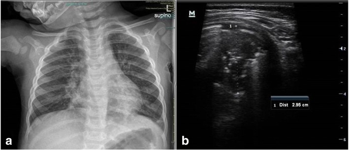
Fig. 3.
Comparison of CXR and LUS in a patient with bronchiolitis complicated by pneumonia in the left lung. a CXR demonstrated a basal left consolidation suggestive of pneumonia and bilateral peri-bronchial thickening. b Ultrasound showed a consolidation with air bronchograms in the posterior region of the left lung

