Fig. 4.
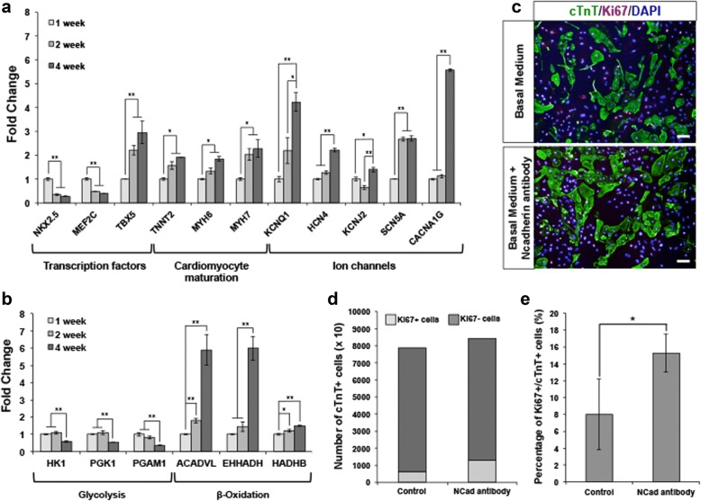
Quantitative PCR analysis of mRNA isolated from cardiomyocytes at various time points post-initial contraction (1, 2, and 4 weeks). Upregulation of Wnt signaling leads to proliferation of human ES cell-derived cardiomyocytes. a The relative gene expression levels showed downregulation of early key transcription factors governing cardiogenesis (NKX2.5 and MEF2C) and upregulation of genes associated with cardiomyocyte maturation such as MYH7, KCNQ1, SCN5A, HCN4, and CACNA1G. b Concomitantly, metabolic shift from glycolysis to β-oxidation was observed. Error bars indicate s.d., n = 3 replicates. *P < 0.05 and **P < 0.01 for Kruskal-Wallis one-way analysis of variance compared to control. c Representative images of untreated control and N-cadherin antibody-treated human ES cell-derived cardiomyocytes immunostained for Ki67 (red) and cTnT (green). Cell nuclei (blue) were stained with DAPI. Scale bar, 50 μm. d, e Graphical quantification of the proportion of Ki67+ cells in N-cadherin antibody-treated human ES cell-derived cardiomyocytes compared to untreated cardiomyocytes (control). There is an increase of ~ 1.0-fold change of Ki67+/cTnT+ cells in N-cadherin antibody-treated cardiomyocytes compared to control. Error bars indicate s.d., n = 3 experiments, *P < 0.05, evaluated by Student’s t test

