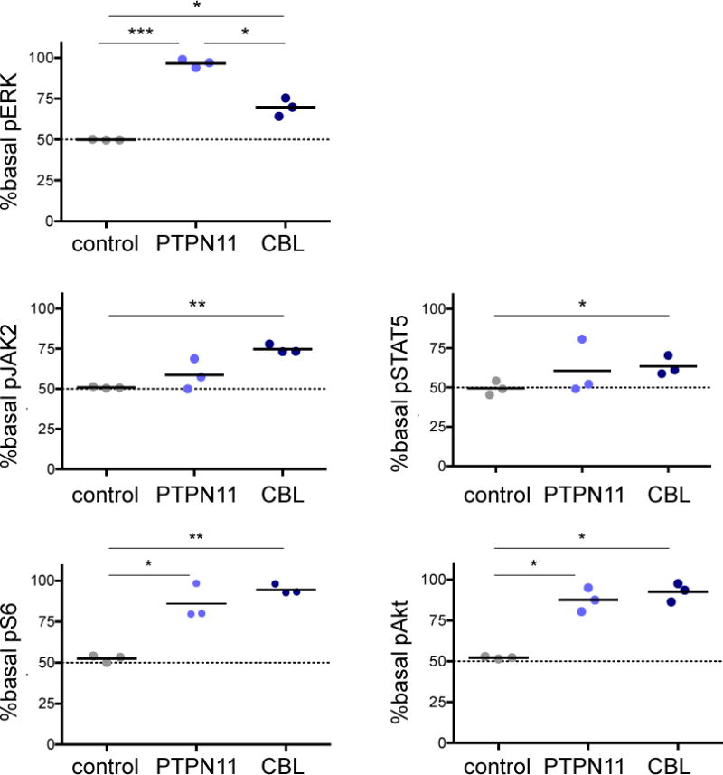
Figure 2. Constitutive signaling activation in PTPN11-mutant and CBL-mutant JMML iPSC-derived myeloid cells.

Phosphoflow cytometric analysis of day 14 CD45+14+18+ myeloid cells from control, PTPN11 and CBL iPSCs was performed. Basal levels of phosphorylated (p) ERK, JAK2, STAT5, S6, and AKT in CD45+14+18+ control or JMML iPSCs were measured and normalized to the median level of each phosphoprotein in control cells for comparison of signaling activation. Data points denote three independent experiments with means (thick black lines). Gating strategy of surface markers and median control phosphoprotein levels is depicted in Supplemental Figure 1. Data were analysed by one-way ANOVA with the Tukey post-test for multiple comparisons. *p<0.05, **p<0.01, ***p<0.001.
