
Figure 1. BD-RGCs axon terminal locations throughout the dLGN.
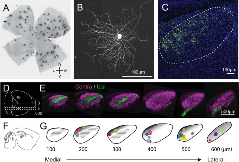
A. Image of densely labeled BD-RGCs in a whole mount retina from the FSTL4-CreERT2 line. Consistent with previous studies, we find that the dorsal retina is more densely labeled than ventral side. B. Single BD-RGC in the retina. For single-cell reconstructions, low-dose tamoxifen was used to label just one or a few cells to ensure isolated arbors. C. dLGN from a mouse with densely labeled BD-RGCs. Axons are found predominantly in the dorsal-caudal region of the nucleus. D. Schematic of dorsal view of sagittal sections in the intact mouse brain (LGNs in magenta and green). E. CTB-labeled retinal axon projections delineate contralateral (magenta) and ipsilateral (green) regions within sagittal sections of the dLGN. F. Schematic of sagittal section with dLGN. G. dLGN template of individual 100 μm sections from medial to lateral aspects of the dLGN with the location of all 26 reconstructed axons. Each arbor’s location is color coded and mapped to the section of the dLGN where it was located. Size and shape of the colored symbols reflect the perimeter of the axon arbor. Gray shaded areas roughly approximate the shell region of the dLGN as previously described in mice for coronal sections (Grubb & Thompson, 2004; Cruz-Martín et al., 2014) and estimated on sections in the sagittal plane. Hatched black regions represent the ipsilateral zone as shown in green in panel E. The distinction between shell and core remains poorly defined at the rostral end. Panels 1C and E are modified from (Hong et al., 2014).