
FIGURE 2.
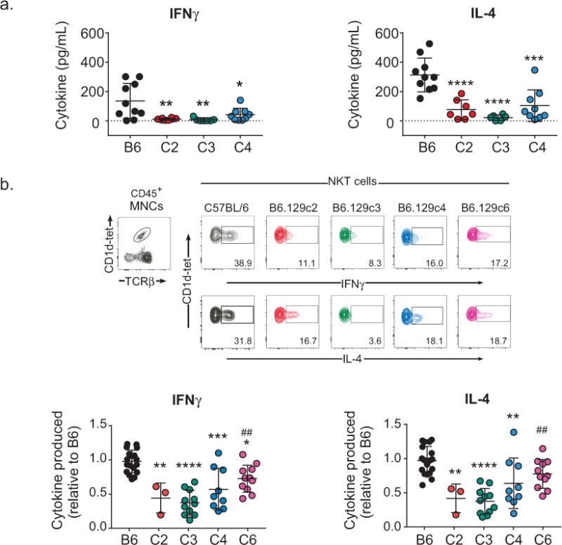
Regulation of αGalCer-induced iNKT cell cytokine production maps to a 0.14 Mbp region on chromosome 1. A). Decreased serum cytokine in B6.129 congenic mice in response to αGalCer. Blood was collected from B6 or B6.129 congenic mice 2 h after αGalCer administration. Serum cytokine levels were assessed using ELISA. Statistical analysis was performed using 1-way ANOVA followed by Sidak’s multiple comparisons test, *p ≤ 0.05, **p ≤ 0.01, ***p ≤ 0.001, ****p ≤ 0.0001. B). Top: Representative intracellular staining of cytokine-producing iNKT cells in response to αGalCer. Splenocytes were isolated 2 h after αGalCer administration. iNKT cells were identified using CD1d-tetramer/PBS57 and TCRβ. The percentages of iNKT cells expressing cytokines were determined using an isotype control for each mouse. Lower: Decreased iNKT cell cytokine production in B6.129 congenic mice in response to αGalCer. The data represent the relative level of iNKT cell cytokine production. Data are the combined data from 5 separate experiments using female mice 8-14wks of age and are presented as the mean ± s.d. All mice were age-matched to controls in each experiment. Statistical analysis was performed using 2-way ANOVA followed by Tukey’s multiple comparison test. * = comparison of B6 to B6.129 congenics. # = comparison of B6.129c3 to B6.129 congenics. *p ≤ 0.05, **, ##p ≤ 0.01, ***p ≤ 0.001, ****p ≤ 0.0001.