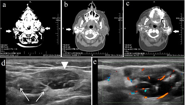
Figure 3.

Imaging features in patient B. Head and neck CT scan: (a) native CT scan in transverse plane shows both parotid glands (arrows) increased in size and displaying structural changes with a cystic-like pattern; (b-c) sialotomography images after administration of contrast agent in which both right (b) and left (c) parotid glands display dilated canalicular ducts. Parotid gland US exam: (e) B-Mode US longitudinal scan image displays severe PIH, with large anechoic areas, some measuring over 1 cm (arrowhead), frequently confluent, multiple cysts and calcifications (arrows), severely modified glandular architecture; (f) Power Doppler US image which shows increased vascularity pattern
