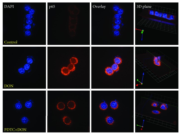
Figure 2.
Nuclear translocation of phosphoryl-NF-κB p65 (p-p65 (Ser536)) induced by DON treatment (1200 ng/mL) and PDTC pretreatment (20 μM, 45 min) followed by DON treatment (1200 ng/mL) in GH3 cells, visualized through indirect immunofluorescence, using Alexa Fluor-conjugated secondary antibody. The nucleus was stained with PI. The panels show PI staining, Alexa Fluor staining, overlay, and the 3D plane of the cells. All photos were captured at 400x magnification. Phosphoryl-NF-κB p65 was upregulated and can be observed in the nucleus.

