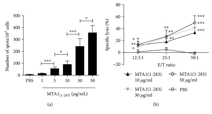
Figure 4.
Specific T cells induced by MTA1(1–283) secrete IFN-γ and lyse target cells. PBMCs from healthy HLA-A∗02+ donors were isolated and stimulated with protein MTA1(1–283) (at final concentrations of 0, 1, 5, 10, 30, and 50 μg/ml) in RPMI 1640 medium supplemented with 50 U/ml interleukin-2 and 10% FCS once a week for 21 days. On day 21, the induced T cells were collected; (a) IFN-γ secretion was assessed by the ELISPOT assay; ∗ P < 0.05 and ∗∗∗ P < 0.001 between two groups. (b) Cytotoxic activity was assessed by the LDH assay using SW620 cells (HLA-A2+, MTA1+) as the target cells. T cells induced by PBS were used as negative controls. ∗ P < 0.05, ∗∗ P < 0.01, and ∗∗∗ P < 0.001 vs. the control group. Data represent means ± SD (n = 4).

