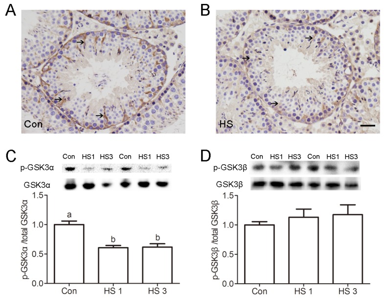
Figure 2.

Heat shock-induced dephosphorylation of GSK3α in Sertoli cells. (A-B) Representative microscopic images of p-GSK3α in control (A) and heat shock (HS) treated (B) mouse testis evaluated by immunohistochemistry. Arrows indicate p-GSK3α-positive spermatocytes. Scale bar=50 μm. (C) Western blots and histogram showing the protein levels of p-GSK3α and GSK3α in mouse testis after heat shock. (D) Western blots and histogram showing the protein levels of p-GSK3β and GSK3β in mouse testis after heat shock. Con, control; HS, heat shock. Values are expressed as the mean±SEM, n=6. Values with different superscripts are significantly different from each other (P<0.05).
