Figure 6.
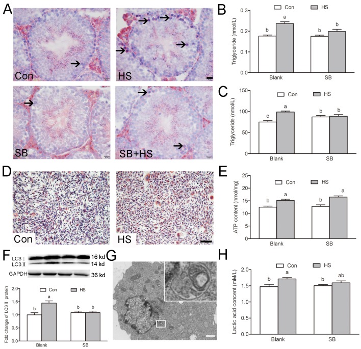
GSK3α activation participates in HS-induced Sertoli cells lipid droplets accumulation. (A) Representative microscopic images of lipid droplet formation in mouse testis treated with HS or GSK3α inhibitor. ORO stained lipid droplets are shown in red (arrows). Scale bars=20 μm. (B) Histogram showing quantification of TG content in mouse testis treated with HS or GSK3α inhibitor. (C) Histogram showing quantification of TG content in TM4 cells treated with HS or GSK3α inhibitor. (D) Representative microscopic images of lipid droplet formation in TM4 cells. Scale bars=50 μm. (E) Histogram showing quantification of ATP content in TM4 cells treated with HS or GSK3α inhibitor. (F) Western blots and histogram showing the protein levels of LC3 in TM4 cells. (G) Representative electron microscopic images of autophagosome structure in TM4 cells. (H) Histogram showing quantification of lactic acid content in TM4 cells treated with HS or GSK3α inhibitor. Con: control; HS: heat shock; SB: SB216763. Values are expressed as the mean±SEM, n=6. Values with different superscripts are significantly different from each other (P<0.05).

