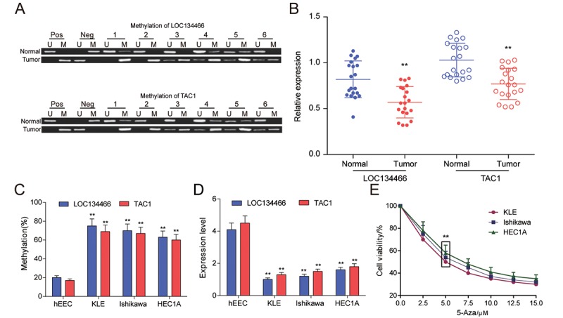
Figure 7.

LOC134466 and TAC1 were hypermethylated and lower-expressed in EC tumors. (A) Methylation state of LOC134466 and TAC1 in tumor tissues and paired adjacent tissues. There were 20 paired tissue samples and six representative MSP results were represented. (B) The expressions of LOC134466 and TAC1 in tumor tissues and paired adjacent tissues were assessed by qRT-PCR. The expression of gene was normalized to that of GADPH. (C) DNA methylation level of LOC134466 and TAC1 in normal endometrial cell line (hEEC) and EC tumour cell lines (KLE, Ishikawa and HEC1A) were determined by MSP. (D) The LOC134466 and TAC1 mRNA expressions in cancer cell lines and normal cell line were analyzed by real-time PCR. The expressions of genes were normalized to that of GADPH. (E) Minimum effective dose of 5'-Aza-deoxycytidine was determined by cell viability assay. 5 μM 5’-Aza-deoxycytidine showed a great difference. ** P<0.01 compared to corresponding control (paired normal tissues or hEEC cell line or cell viability without 5’-Aza treatment).
