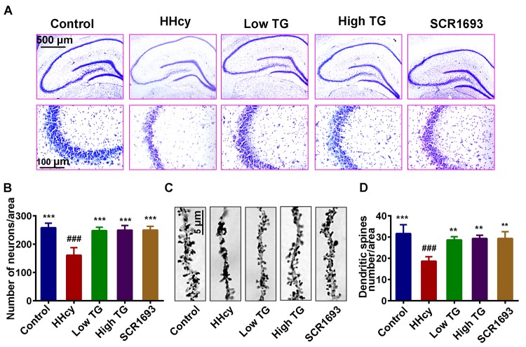
Figure 3.
Supplementation with TG recovered dendritic spine and neuronal loss. After two weeks of Hcy (400 µg/kg/day) and two weeks TG treatment post-injection, the rats were sacrificed following behavioral test. (A and B) Representative Nissl staining images and the quantification of neuronal density, chart bar = 500 and 100 µm for low and high magnifications respectively. (C and D) Representative Golgi staining images and quantification of dendritic spines from randomly selected dendritic segments of randomly selected hippocampal neurons, chat bar = 5µm. HHcy animals showed neurodegeneration which was recovered following supplementation with both low and high TG doses. The data were expressed as mean ± SEM and n = 3 for both Nissl and Golgi staining. ### P < 0.001 versus control; ** P < 0.01, *** P < 0.001 versus HHcy.

