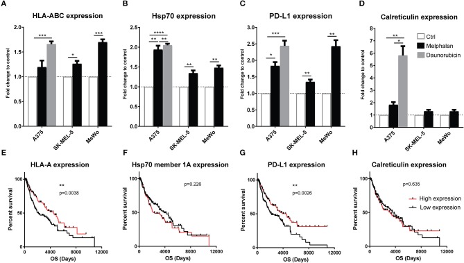
Figure 3.
Melanoma cells exposed to melphalan upregulate immune-related surface markers. The human melanoma cell lines A375, SK-MEL-5 and MeWo were exposed to sub-lethal concentrations of melphalan for 1 h at 40°C (n = 6 for all cell lines). The melphalan was then washed away and the cells were cultured for an additional 24 h at 37°C. A375 cells were also exposed to a sub-lethal concentration of daunorubicin for 24 h (n = 5). The expression of (A) HLA-ABC, (B) Hsp70, (C) PD-L1, and (D) calreticulin was then determined by flow cytometry. The data show fold change in expression compared with non-exposed cell controls on the living populations. Paired t-test. Data are presented as mean values with SEM. Melanoma patients in the TCGA database were dichotomized into two groups based on above or below median gene expression of (E) HLA-A, (F) Hsp70 member 1a, (G) PD-L1, and (H) calreticulin, followed by analysis of overall survival (OS) by the log-rank test. *P ≤ 0.05, **P ≤ 0.01, ***P ≤ 0.001, ****P ≤ 0.0001.

