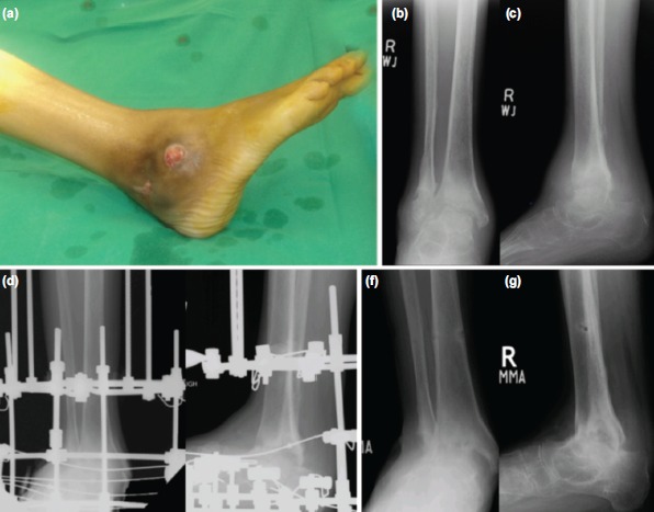
Fig. 1:

(a) Intra-operative photograph of 45 year-old lady with tuberculosis ankle joint showing ulcers at ankle. (b,c) Pre-operative radiographs Ilizarov anteroposterior and lateral views showing subchondral erosions and sclerosis. (d,e) Immediate postoperative radiographs showing Ilizarov fixator across ankle joint Ilizarov anteroposterior and lateral views. (f,g) Nine months post-operative radiographs Ilizarov anteroposterior and lateral views after removal of Ilizarov fixator showing ankle joint fusion.
