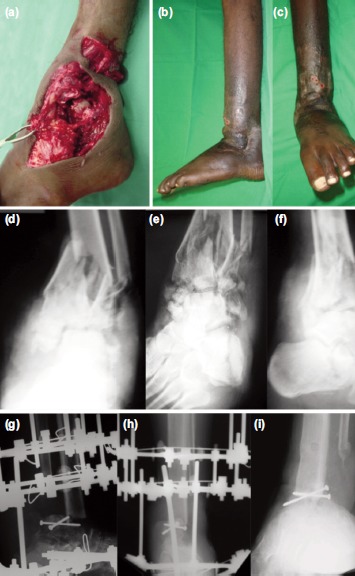
Fig. 3:

(a) Intra-operative photographs showing comminuted fracture left ankle. (b,c) Photographs after ankle fusion and removal of Ilizarov. (d,e) Pre-operative radiographs anteroposterior, mortis and lateral views showing comminuted fracture left ankle. (f,g) Immediate postoperative radiographs showing ankle arthrodesis with Ilizarov anteroposterior, lateral views. (h,i) Fifteen months post-operative radiographs showing fusion Ilizarov anteroposterior, lateral views.
