Figure 2.
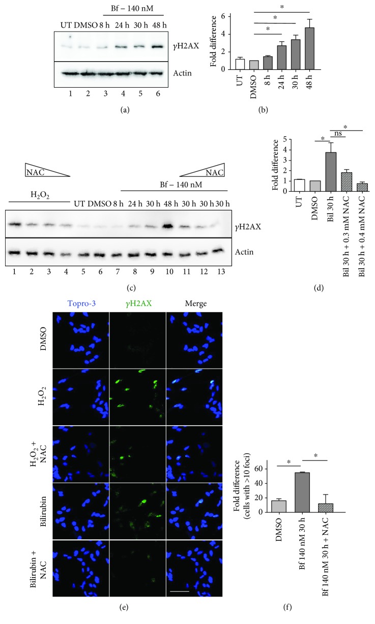
Bilirubin-induced oxidative stress causes DNA damage in SH-SY5Y cells. SH-SY5Y cells were treated with 140 nM free bilirubin (Bf) for different time points (8, 24, 30, and 48 h; lanes 3-6). DMSO (0.6% DMSO, bilirubin solvent) served as controls (lane 2). Cells were collected at the indicated time points, and the levels of γH2AX were determined by Western blot analysis and quantified (mean of three independent experiments (b)); (c, d) SH-SY5Y cells were treated with H2O2 (lanes 1-4) or 140 nM Bf for different times, as indicated (lanes 7-13). In lanes 2-4 and 11-13, cells were treated with H2O2 or 140 nM Bf for 48 h plus N-acetyl-cysteine (NAC) treatment (0.1, 0.3, or 0.4 mM). γH2AX was determined by Western blot, quantified and normalized (actin). (d) Quantification of two independent experiments; (e, f) SH-SY5Y cells were treated with H2O2 or 140 nM bilirubin for 30 h, with or without NAC (0.4 mM) treatment, as described in Materials and Methods. γH2AX foci were determined by immunofluorescence. Cells containing more than 10 foci were considered positive. The scale bar corresponds to 30 μM; (f) quantification from two independent experiments. Values represented as mean ± SD. One-way ANOVA with Bonferroni's post hoc test was applied for statistical analysis. ∗ P < 0.05; NS: not significative; UT: untreated cells.

