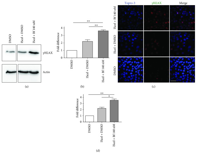
Figure 3.
Bilirubin-induced DNA damage in HeLa DR-GFP cells. (a, b) Western blot analysis of HeLa DR-GFP cells transfected with a plasmid encoding the ISceI endonuclease (pCBA IsceI) and treated with DMSO (control) or 140 nM free bilirubin (Bf). γH2AX levels were determined by Western blot analysis, quantified and normalized (actin). (b) Quantification from three independent experiments. (c, d) HeLa DR-GFP cells were treated as described in (a) and (b), and γH2AX foci were determined by immunofluorescence. Cells containing more than 10 foci were considered positive. (f) Quantification from two independent experiments. One-way ANOVA with Bonferroni's post hoc test was used to perform statistical analysis. ∗ P < 0.05; ∗∗ P < 0.01.

