FIGURE 2.

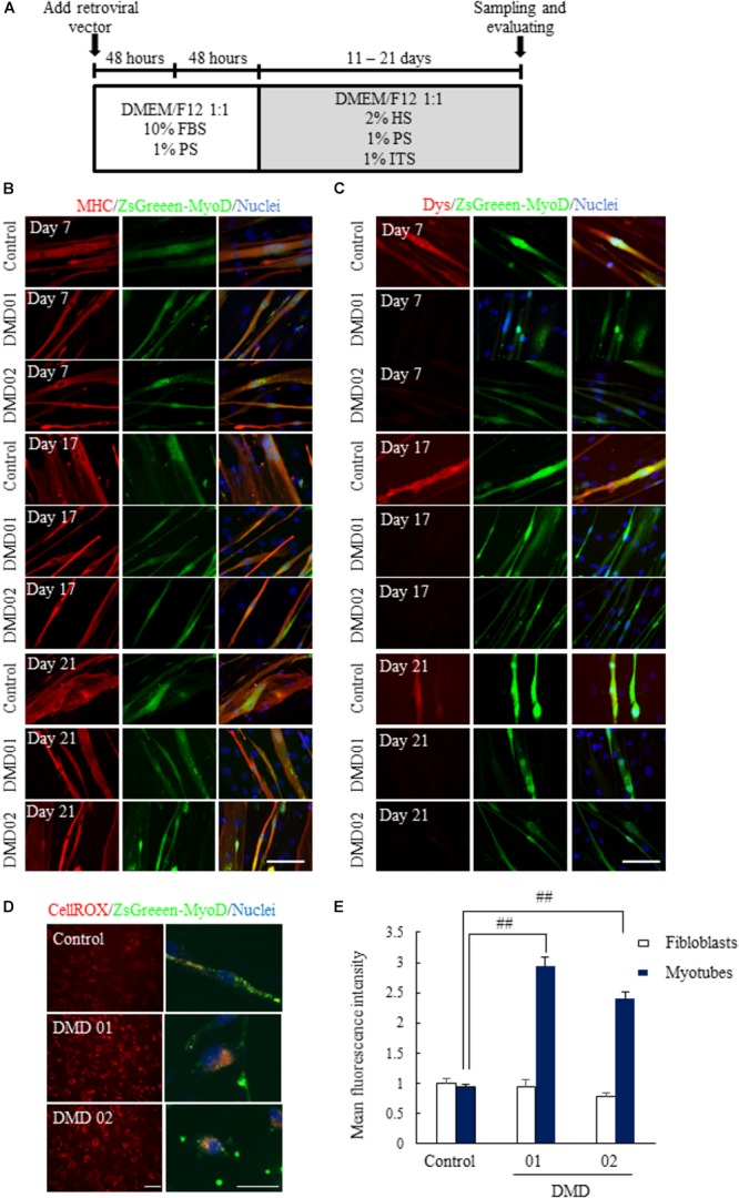
Conversion of fibroblasts to myotubes by MyoD transduction. (A) Schematic of myotubes differentiation from MyoD-transduced fibroblasts. (B) Immunostaining of skeletal muscle marker (myosin heavy chain, MHC) 11 to 21 days after myogenic differentiation. (C) Immunostaining of dystrophin 11 to 21 days after myogenic differentiation. The myotubes from MyoD-transduced fibroblasts of DMD patient resulted in the decrease of dystrophin protein. Scale bar = 50 μm. (D,E) ROS assay using CellROX® deep red reagent to investigate whether ROS production was promoted or not. Data are shown as means ± SEM (n = 5). ##p < 0.01 vs. Control (Student’s t-test). Scale bar = 100 μm (left image), 50 μm (Right magnified image).
