
FIGURE 3.
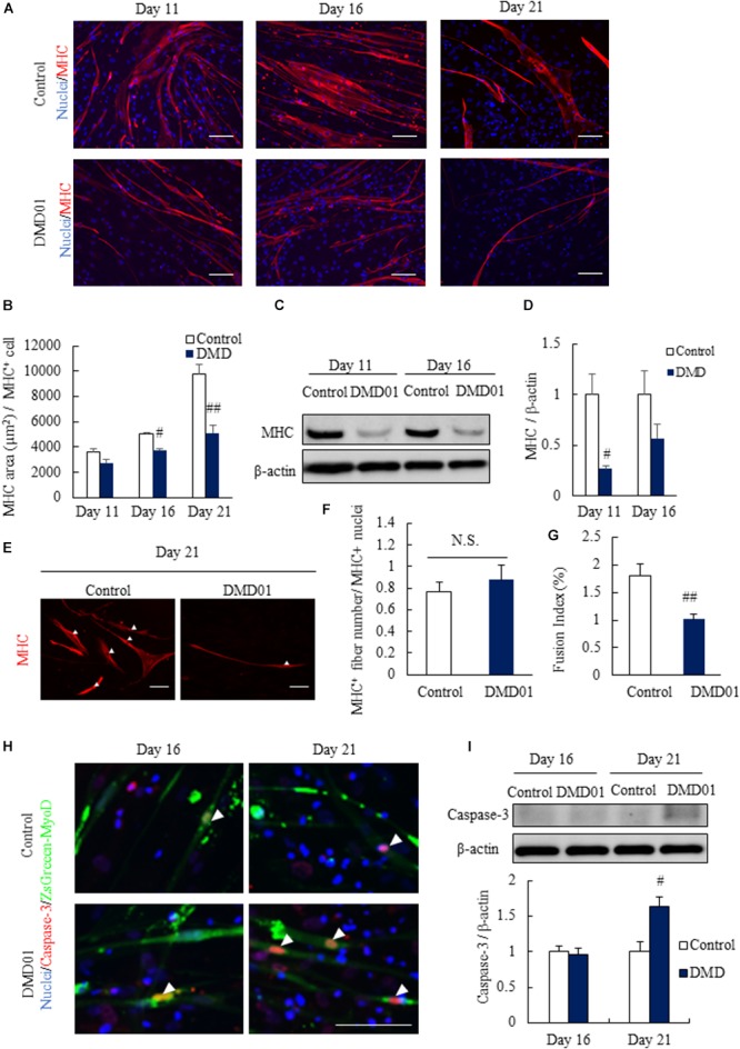
Comparison between myotues from MyoD-transduced fibroblasts of a normal human cell line and a DMD patient. (A) Comparison of myotubes from MyoD-transduced fibroblasts of a control individual and patient with DMD 11, 16, and 21 days after myogenic differentiation using skeletal muscle marker (myosin heavy chain, MHC). (B) Quantification of myotube area in MyoD-transduced fibroblasts of a control individual and a patient with DMD 11, 16, and 21 days after myogenic differentiation by analyzing MHC positive cells. Data are shown as means ± SEM (n = 3 or 5). ##p < 0.01 and #p < 0.05 vs. Control (Student’s t-test). (C,D) Western blot analysis of MHC in a Ctrl individual and a DMD patient derived myotubes at 11 and 16 days. Data are shown as means ± SEM (n = 3). #p < 0.05 vs. Control (Student’s t-test). (E,F) Number of nuclei per MHC+ myotubes in Ctrl individual and a DMD patient at 21 days. Arrowheads indicate the nuclei. Data are shown as means ± SEM (n = 5). Scale bar = 100 μm. (G) Fusion index analysis in Ctrl individual and a DMD patient (DMD01). Data are shown as means ± SEM (n = 3). ##p < 0.01 vs. Control (Student’s t-test). (H) Comparison of death of myotubes from MyoD-transduced fibroblasts of a control individual and a patient with DMD 16 and 21 days after myogenic differentiation using apoptosis marker (cleaved caspase-3). Arrowheads indicate cleaved caspase-3 positive cells. Scale bar = 100 μm. (I) Western blot analysis of cleaved caspase-3 in myotubes from MyoD-transduced fibroblasts of a control individual and a DMD patient. Data are shown as means ± SEM (n = 3). ##p < 0.01 vs. Control and #p < 0.05 vs. Control (Student’s t-test).